Congenital Heart Disease

Congenital heart disease (CHD) is the term used for any abnormality of the heart due to faulty development before birth. With an incidence of about 1% of live births, CHD is one of the most important and common congenital malformation and the leading cause of birth-defect related morbidity and mortality especially during infancy. Heart defects originate in the early weeks of pregnancy when the heart is forming. Not all cases of CHD take the form of “hole in the heart”. Practically any component of the normal heart can be abnormally formed.
What Causes CHD?
Most CHDs are due to a combination of genetic and environmental factors during pregnancy. Other causes that can account for a small number of patients include rubella infection during pregnancy, genetic syndromes like Down’s anomaly (where half of the babies have CHDs), consumption of certain medications during pregnancy, and some rare genetic disease. Oftentimes, it is not possible to identify the specific cause.
Heredity may play a role in some heart defects. A parent who has a CHD is slightly more likely than others to have a child with the problem (risk of about 1 in 30) and more than 1 child in a family can be born with a heart defect (risk of about 1 in 50).
The news that your child has a CHD will probably make you anxious and worried about your child’s immediate and long-term health. Rest assured that most CHDs are simple, can get better on their own or eventually heal and themajority do not need treatment. Only a quarter of babies with CHD require some form of treatment in the first year of life.
What is “Hole in the Heart”?
A hole in the heart is a type of simple CHD that changes the normal flow of blood through the heart. Most babies are born with a single small hole.
The normal heart has 2 sides, separated by an inner wall called the septum, and 2 upper chambers, the right (RA) & left atrium (LA) and 2 lower chambers, the right (RV) & left ventricle (LV).The right heart receives oxygen-poor blood from the body and pump it to the lungs. The left side of the heart receives oxygen-rich blood from the lungs and pumps it to the body. The septum prevents mixing of blood between the two sides of the heart.
The normal heart has 4 valves – mitral (between LA & LV), tricuspid (between RA & RV), pulmonary (outflow to lungs) and aortic valve (outflow to body).
A hole in the septum between the heart’s upper chambers is called an atrial septal defect (ASD) and one in the septum between the heart’s 2 lower chambers is called a ventricular septal defect (VSD). An infant born with ASD or VSD may have a single hole, more than one hole or a combination of the two types. Other anomalies can co-exist such as problems with the heart valves and blood vessels of the heart.
ASDs and VSDs allow blood to pass from the left side of the heart to the right side, causing oxygen-rich blood to be pumped to the lungs a second time.
For more details on Artial Septal Defect and Ventricular Septal Defect, go to The New Age Parents Feb/March 2012 issue
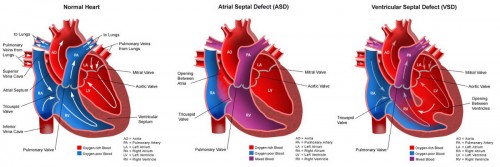 [Click on the image for a bigger picture]
How is a hole in the heart diagnosed ?
Holes in the heart are usually diagnosed based on clinical signs from physical examination and special tests. The diagnosis can be made from presence of a heart murmur, rhythm irregularity, effort intolerance, failure to thrive, or incidental discovery of an abnormal ECG and CXR. They can also be detected during pregnancy from an abnormal fetal heart scan.
In some, physical signs may not be apparent and the diagnosis may not be made until later childhood or even adulthood.
The electrocardiograph (ECG) detects and records electrical activity of the heart by showing how fast the heart is beating, whether the heart rhythm is steady or irregular and give an indication of any heart muscle stress. Chest X-ray shows the size & shape of the heart and whether the lungs have extra blood flow or fluid which can be a sign of heart failure.
Confirmation of the structural defect is carried out by ultrasound examination which uses sound waves to create a moving picture of the heart. In echocardiography, a complete anatomical survey of the malformed heart is worked out from cross-sectional imaging. This allows the cardiologist to clearly examine any problem with the way the heart is formed or the way it’s working, and how the heart is reacting to these defects, and guiding whether and when treatment is needed.
Cardiac catheterizationis carried out under general anaesthesia to provide additional information on special cardiac structures and provide information on oxygen content and blood pressure profile of the heart. A thin flexible tube or catheter is put into a vein in the groin and threaded into the heart. A radio-opaque substance (dye or contrast) that can be seen on X-rays is then injected through the catheter into a blood vessel or a chamber of the heart forming X-ray images of the heart structures.

How is a hole in the heart treated ?
Many holes do not need treatment. More than half of ASDs &VSDs eventually close or get smaller. Management depends on the type, location, and size of the hole/s. Other factors include the age, weight and general health. Medications are given to help the heart work better when there is heart failure and heart rhythm abnormalities. The hole/s can be repaired by open heart bypass surgery or device closure via an interventional catheterization. Closure of medium to large ASDs are done by the time the child is 2 to 5 years of age.
Trans-catheter closure of holes in the heart with septaloccludershave been available in Singapore since 1997. This method can be performed on most secundum ASDs and can also be done for certain types of VSDs. An umbrella-like device is folded up and pushed out of the catheter and positioned so that it plugs the hole between the atria. Within 6 months, normal tissue grows in and over the device. Catheter procedures are much easier than surgery because they involve only a needle puncture in the groin where the catheter is inserted. Recovery is faster and easier, with no scar. In the right candidates, closures are successful in 9 out of 10 patients, with no significant residual leakage.
Some defects can increase the risk of a serious infection of the heart valves or the inner surfaces of the heart (infective endocarditis). Prevention of this by appropriate antibiotic prophylaxis is important before surgery or dental procedures that could allow bacteria to enter the blood stream and cause endocarditis.
Living with a hole in the heart
Many children do not need any special care, active intervention or treatment except for regular checks with a cardiologist. There is no activity restrictions on those with small defects.The general rule is that babies and children should be treated as far as possible, as if they are normal. All the usual immunisations and vaccinations should be done and at the recommended scheduled time. The outlook for children with ASDs and VSDs is excellent. Early and accurate diagnosis with advances in treatment and low risk of surgery mean that most children with these heart defects have normal, active and productive lives with no decrease in lifespan.
Dr Chan Kit Yee, Paediatrician, Special Interest in Cardiology.
Dr Chan graduated from the National University of Singapore with a Bachelor of Medicine and Bachelor of Surgery degree in 1981 and obtained her Masters of Medicine (Paediatrics), Singapore, in 1986. She was conferred a Doctor of Medicine (M.D.) degree by NUS for her thesis on cardiac dysrhythmias in children in 1996.
Practice Address:
SBCC Baby & Child Clinic (Mount Alvernia)
820 Thomson Road, Mount Alvernia Medical Centre Blk A
#01-01/02 Mount Alvernia Hospital
Singapore 574623
Tel: 6354 1922
This article was first published in The New Age Parents e-magazine.
* * * * *
Like what you see here? Get parenting tips and stories straight to your inbox! Join our mailing list here.
Want to be heard 👂 and seen 👀 by over 100,000 parents in Singapore? We can help! Leave your contact here and we’ll be in touch.












Leave a Comment: